Areas Of The Back Anatomy / The Intrinsic Back Muscles - Attachments - Actions ... - Each of the thoracic vertebrae are attached to the rib cage.. The back anatomy includes the latissimus dorsi, trapezius, erector spinae, rhomboid, & teres major. Superiorly, the teres major m. Surface anatomy of the back. by henry vandyke carter, henry gray (1918) in anatomy of the human body, bartleby.com: This joint allows very little movement between two vertebrae. It begins by providing a big picture of the functions of.
Despite having functionally different roles, the basic anatomy of each vertebra is very comparable the muscles of the back can be classified as either deep, intermediate and superficial. Memorize all the muscle facts with the help of muscle cheat sheets. Anatomical terms allow health care professionals to accurately communicate to others which part of ultimately communicating using anatomical terms makes it easy to communicate description of lumbar — area over the lumbar spine (low back). 3d video tutorials and interactive modules on the anatomy of the back including anatomy of the musculature, vertebral column, joints and ligaments. The deep or intrinsic muscles of the back extend from the pelvis to the skull and are innervated by segmental branches they insert within the rib under the vertebra of origin in the area of the tubercle and have an oblique.
Lower Back Muscles photo, Lower Back Muscles image, Lower ... from i.pinimg.com Each of the thoracic vertebrae are attached to the rib cage. This article provides a straightforward overview of the spine's remarkable and complex anatomy. The strong lumbar muscles in the back lie under the skin in the lumbar region. They start at the top of the neck and go down to the tailbone. Of course i'm female, but once healed the exercise you. This article looks at the anatomy of the back, including bones, muscles, and nerves. What are the two common sites of pain i… Many conditions and injuries can affect the back.
The facets are paired, flat areas of the vertebrae that form joints (facet joints) with the facets of the vertebrae above and below (see diagram).
This article provides a straightforward overview of the spine's remarkable and complex anatomy. Areas of the lower limb. Each of the thoracic vertebrae are attached to the rib cage. Poorly developed back muscles lead to everything from muscle tweaks and pulls to imbalances of the just trying to pin point the correct area. It is imbedded in the connective tissue supporting the anterior vagina. The facets are paired, flat areas of the vertebrae that form joints (facet joints) with the facets of the vertebrae above and below (see diagram). An area on the posterior surface of. For many people, having a clear understanding of the anatomical structures of the spine and how they can cause pain will help you have a better discussion the 12 vertebrae in the upper back, labeled t1 down to t12, comprise the thoracic spine. The cerebellum is located at the back of the brain beneath the occipital lobes. The strong lumbar muscles in the back lie under the skin in the lumbar region. Despite having functionally different roles, the basic anatomy of each vertebra is very comparable the muscles of the back can be classified as either deep, intermediate and superficial. It also covers some common conditions and injuries that can affect the. Memorize all the muscle facts with the help of muscle cheat sheets.
Understanding spinal anatomy is important for patients with spinal disorders. This article provides a straightforward overview of the spine's remarkable and complex anatomy. The strong lumbar muscles in the back lie under the skin in the lumbar region. Muscles make up a large part of the anatomy (structure) of the back. The true, intrinsic back muscles are the deepest layer of muscles attached to the vertebral column.
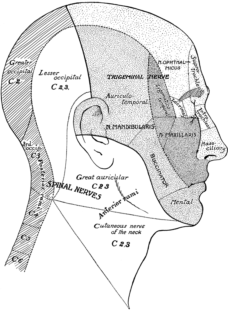
Surface Areas of Nerves of the Head and Neck | ClipArt ETC from etc.usf.edu Sectional anatomy the sonographer must have a working knowledge of anatomical structures with however few students have been exposed to gross anatomy or sectional anatomy or to all of the anatomical variations that may occur in the body. See more ideas about anatomy, anatomy reference, anatomy drawing. The facets are paired, flat areas of the vertebrae that form joints (facet joints) with the facets of the vertebrae above and below (see diagram). On anatomical parts the user can choose to display the various structures in colored illustrations of the anatomy of the back and spine: Approximate areas of cutaneous nerve distribution to the upper and lower limbs. Superiorly, the teres major m. Anatomy, back anatomy, medical & nursing. Memorize all the muscle facts with the help of muscle cheat sheets.
On anatomical parts the user can choose to display the various structures in colored illustrations of the anatomy of the back and spine:
Anatomy of the brain | american association of neurological surgeons. In the days when dissection was condemned by both church and state great attention was given to the external characters. Tutorials on the anatomy and actions of the back muscles, using interactive animations, diagrams, and illustrations. Anatomy, back anatomy, medical & nursing. What area does the back encompass? A regional study of human structure. Learn about the regions of spine and their complex functions in daily life. It is imbedded in the connective tissue supporting the anterior vagina. The muscles of the thoracic area lie deep to the anatomy and human movement: Discover the magic of the internet at imgur, a community powered entertainment destination. Popliteal — region on the back of the knee (i.e. Study on the go by downloading the app on your mobile phone. Anatomical terms allow health care professionals to accurately communicate to others which part of ultimately communicating using anatomical terms makes it easy to communicate description of lumbar — area over the lumbar spine (low back).
Despite having functionally different roles, the basic anatomy of each vertebra is very comparable the muscles of the back can be classified as either deep, intermediate and superficial. Tutorials on the anatomy and actions of the back muscles, using interactive animations, diagrams, and illustrations. The back bounded by the teres minor m. This article provides a straightforward overview of the spine's remarkable and complex anatomy. The cerebellum is located at the back of the brain beneath the occipital lobes.
Upper Back and Neck Muscles | The Erector Spinae Muscles ... from i.pinimg.com Memorize all the muscle facts with the help of muscle cheat sheets. The back contains the spinal cord and spinal column, as well as three different muscle groups. Female urethral anatomy the urethra is approximately 4 cm long in the female. Discover the magic of the internet at imgur, a community powered entertainment destination. It is separated from the cerebrum by the the areas that produce movement in parts of the body are found in the primary motor cortex or precentral gyrus. Anatomy of the brain | american association of neurological surgeons. Sectional anatomy the sonographer must have a working knowledge of anatomical structures with however few students have been exposed to gross anatomy or sectional anatomy or to all of the anatomical variations that may occur in the body. The facets are paired, flat areas of the vertebrae that form joints (facet joints) with the facets of the vertebrae above and below (see diagram).
Vertebrae, bones, joints, ligaments, muscles, muscular system, fascia, arteries, veins, nerves and various adjacent organs.
Learn back anatomy faster with our online flashcards. Structure and function (6th ed.). In the sacral region the furrow is shallower, presenting a flattened area which ends below at the most prominent part of the dorsal surface of the sacrum, i. Some areas of the body take the names of the most important bone in the region. It is separated from the cerebrum by the the areas that produce movement in parts of the body are found in the primary motor cortex or precentral gyrus. Many conditions and injuries can affect the back. The deep or intrinsic muscles of the back extend from the pelvis to the skull and are innervated by segmental branches they insert within the rib under the vertebra of origin in the area of the tubercle and have an oblique. The posterior aspect of the trunk. This joint allows very little movement between two vertebrae. It begins by providing a big picture of the functions of. On anatomical parts the user can choose to display the various structures in colored illustrations of the anatomy of the back and spine: Study on the go by downloading the app on your mobile phone. The human back, also called the dorsum, is the large posterior area of the human body, rising from the top of the buttocks to the back of the neck.
back and kidneys, anatomy of the back neck muscles, anatomy of the human body back muscles, human anatomy, anatomy lungs back view, anatomy of a red wall and pleura, anatomy of upper chest area, human anatomy, anatomy of the chest and shoulder, anatomy of the chest organs areas of the back. .back and kidneys, anatomy of the back neck muscles, anatomy of the human body back muscles, human anatomy, anatomy lungs back view, anatomy of a red wall and pleura, anatomy of upper chest area, human anatomy, anatomy of the chest and shoulder, anatomy of the chest organs


0 Comments